The muscles which are required for mastication or chewing are known as the muscles of mastication. These muscles help mainly in the movement of the mandible and not the maxilla as maxilla is an integral part of the skull and the mandible being the only movable bone in the skull. The muscles help in moving the mandible at the Temporomandibular Joint which helps in chewing or food, talking, yawning, etc.
There are many muscles which help in the process of mastication but the main muscles which take part in the process are
Primary Muscles Of Mastication:
- Masseter muscle
- Temporalis
- Lateral Pterygoid
- Medial Pterygoid
Accessory Muscles Of Mastication:
The secondary muscles or Accessory muscles are divided further into two types based on their position.
Buccinator
Suprahyoid Muscles
- Digastric
- Stylohyoid
- Mylohyoid
- Geniohyoid
Infrahyoid muscles
- Sternohyoid
- Thyrohyoid
- Omohyoid

Courtesy Netter’s Atlas
Now let us discuss in detail about each of these muscles:
Masseter Muscle:
It is one of the main muscle which helps in the process of mastication
In humans, the masseter is the second most efficient masticatory muscle. Its origin and insertion make it very useful for the movement of the jaw and for applying good bite force for mastication.
Masseter muscle is a powerful muscle because of its Multipennate arrangement of fibers
The masseter muscle extends from the zygomatic arch to the ramus and body of the mandible. The fibers of this muscle are broad, extending from the region of the second molar on the surface of the mandible to the surface of the ramus. The muscle is divided into 2 parts
- Superficial
- Deep
Origin of masseter muscle:
- Superficial layer – anterior 2/3rd of lower border of zygomatic arch & zygomatic processof maxilla
- Middle layer – anterior 2/3rd of deep surface & posterior 1/3rd of lower border of zygomatic arch
- Deep layer – deep surface of zygomatic arch
Insertion of masseter muscle:
- Superficial layer –lower part of lateral surface of ramus of mandible
- Middle layer –middle part of ramus
- Deep layer – upper part of the ramus & coronoid process
The main function of masseter muscle is
- Elevation of the mandible
- lateral movements of the mandible for efficient chewing and grinding of the food
- unilateral chewing
- Retraction of the mandible
Blood supply of masseter muscle:
- Masseteric artery .
Nerve supply of masseter muscle:
- Massetric nerve.
Clinical Importance of Masseter Muscle of Mastication:
- Masseter muscle can be palpated both intraorally and extra orally
- Most common muscle involved in Myositis Ossificans
- Masseter Muscle shows Dual action in Complete Denture
- The muscle that commonly undergoes Hypertrophy in Bruxism is Masseter
- Because of the Multipennate arrangement of fibers masseter is a very powerful muscle

Masseter Muscle Function – Elevation of Mandible
Temporalis Muscle:
This is the muscle which helps in elevation of the mandible, It is one of the muscles of mastication. It is large shaped in appearance and covers the Temporal area of the skull.
Origin and Insertion of Temporalis Muscle:
- From the Parietal bone of the skull and is inserted on the coronoid process of the mandible.
Arterial supply of Temporalis Muscle:
- The Deep Temporal artery supplies the large muscle.
Nerve Supply of Temporalis Muscle:
- Trigeminal nerve( this nerve has been associated with being the cause of Headache and migraine.
Functions of Temporalis Muscle:
- Elevation of the mandible
- Retraction of the mandible.
- Crushing of food between the molars.
-
Posterior fibers draw the mandible backwards after it has been protruded.
-
It is also a contributor to side to side grinding movement.
Clinical Importance of Temporalis Muscle:
- Sudden contraction of temporalis muscle will result in coronoid fracture, which is rare.

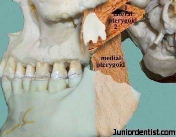
Lateral Pterygoid Muscle:
This is a small muscle which also helps in the mastication process. It is divided into 2 heads
Origin of Lateral Pterygoid Muscle:
- Upper head – infratemporal surface & crest of greater wing of sphenoid bone
- Lower head – lateral pterygoid plate
Insertion of Lateral Pterygoid Muscle:
- Pterygoid fovea on the anterior surface of neck of mandible
- Anterior margin of articular disc & capsule of TMJ
Nerve Supply of Lateral Pterygoid Muscle:
- Pterygoid branch of Trigeminal nerve.
Arterial supply of Lateral Pterygoid Muscle:
- Pterygoid branch of Maxillary artery.
Functions of Lateral Pterygoid Muscle:
- Depresses the mandible
- Protrudes it forward for opening of the jaw
- Side Movements
Clinical Importance of Lateral Pterygoid Muscle:
- Most commonly involved muscle in MPDS
- Only muscle of mastication which has its attachment to the TMJ
- Lateral Pterygoid forms the roof of the Pterygomandibular space.

The combined efforts of the Digastric and Lateral Pterygoid provide for natural jaw opening.
Medial Pterygoid muscle:
It is a thick muscle of mastication.
Origin and Insertion of Medial Pterygoid Muscle:
- It Arises from the deep head the lateral pterygoid plate, and from the maxillary tuberosity.
- Insertion is seen on the Medial angle of the Mandible.
Arterial supplyof Medial Pterygoid Muscle:
- Pterygoid branch of Maxillary artery.
Nerve Supply of Medial Pterygoid Muscle:
- Mandibular nerve through the medial pterygoid.
Functions of Medial Pterygoid Muscle:
- Elevates the mandible,
- Closes the jaw,
- Helps in side to side movement.
Clinical Importance of Medial Pterygoid Muscle:
- Medial Pterygoid muscle can be palpated only intraorally
- Most commonly involved in MPDS
- Trismus following inferior alveolar nerve block is mostly due to involvement of medial pterygoid muscle
Unique features of Masticatory Muscles:
- Have shorter contraction times than most other body muscles
- Incorporate more of muscle spindles to monitor their activity
- Do not have golgi tendon organs to monitor tension
- Elevators predominantly white fibrous which perform fast twitching
- Do not get fatigued easily
- Psychological stress increases the activity of jaw closing muscles
- Occlusal interferences cause a hypertonic synchronous muscle activity
- Closing movement also determined by the height of the teeth
I am really glad that it was helpful to everyone, do let me know if you want any other topics to be explained in a similar way …
What is MPDS? Also, is there anything else unique about the pterygoid muscles?
THanks! PLease also try to email your response, not sure if I can find this site again!
What is MPDS? Also, is there anything else unique about the pterygoid muscles?
THanks! PLease also try to email your response, not sure if I can find this site again!
Thanks a ton!!
My lecturer was very impressed seeing my answer in the exam.
Thanks it’s really helpful
Very clear & precised description
Plzz send me a link so that I can constantly look here for my problematic topics
Nicely explained
Once again thank u so much
Clinical importance part is so helpfull
What sensor nerve innervites the masticator muscules?
Each of these primary muscles of mastication is paired, with each side of the mandible possessing one of the four.
What is MPSD
Please
It is expanded to Myofunctional Pain Dysfunction Syndrome. As the name suggests it is a pain disorder, where certain points on the muscles of the head and neck when triggered lead to excruciating pain. It is mainly Unilateral (on one side of the face only).
THANK YOU SOOOOOOOOOOOO MUCH
THIS POST WAS OF GREAT HELP…..
Very nicely explained.
could u explain why masseter muscle is more sensitive than temporal muscle, when u palpate the muscle especially after long duration mastication activity..? thx
Hi Aditya,
The answer lies in the function of both the muscles, Masseter muscle as the name suggests plays a major role in Chewing food with your Molars of posterior teeth which requires more force and tension. The Temporalis muscle on the other hand, being larger in size and also having more surface area of attachment is only used to elevate or lift the mandible and does not put any force for chewing.
So after a long duration of mastication, the masseter muscle which is smaller and applies more force on the mandible helping to chew any hard substances is more fatigued as compared to Temporalis muscle which only lifts the mandible.
Your notes are incredible ?
Which dual function is performed by massetar in CD