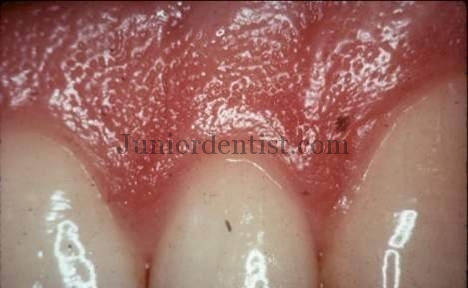
Stippling of Gingiva: Healthy gingiva presents a textured surface like that seen on the surface of an Orange Peel which is called as Stippled. It is also called as Alternate protuberances & depressions on gingival surface.
Stippling was thought to indicate good gingival health, but it has since been shown that smooth gingiva is not an indication of disease, unless it is smooth due to loss of previously existing stippling. Stippling is absent in Infants but it starts to appears in Children from the age of 4.
Stippling is a consequence of the microscopic elevations and depressions of the surface of the gingival tissue due to the connective tissue projections within the tissue.
How and When can Stippling of Gingiva be seen ?
Stippling of Gingiva can best be seen on “Drying the Gingiva”
Which Type of Gingiva is Stippled and which is not ?
- Attached Gingiva – Stippled
- Interdental Gingiva – Stippled
- Marginal Gingiva – Not Stippled
Attached Gingiva is bound to underlying alveolar bone, not the freely movable alveolar mucosa.
Where is Stippling seen Microscopically ?
- Stippling is seen at the sites of fusion of the epithelial ridges or Rete Pegs and corresponds to the fusion of the valleys created by the connective tissue papillae
- It is Microscopic elevations and Depressions of the surface of the gingival tissue due to the connective tissue projections within the tissue.
- The Degree of Keratinization and Stippling are related and depend on each other.
Gingiva comprises of two parts – Free Gingiva and Attached Gingiva. While Free gingiva surrounds the tooth creating a collar around the crown, it is portion of the gingiva which extends from the attached gingiva on to the surface of the tooth. The attached gingiva extends from the free gingiva coronal to the alveolar mucosa in the apical portion of the tooth. Stippling is usually seen in attached gingiva as it is firmly attached to the underlying cementum and alveolar bone with the help of collagen fibers of the connective tissue. Stippling is lost as age progress, in most adult patients above 50 years there is no stippling of Gingiva.
References:
https://www.researchgate.net/publication/329094030_Prevalence_of_Gingival_Stippling_among_4-8_Years_Old_Children
At which particular age stippling is mostly present
Stippling is absent in infancy or new borns and slowly starts to show after a couple of years and starts to increase as age progresses. Stippling starts to disappear in old age. So the most stippling is seen in the middle age group of 20-35 years.
In which light do we see stippling and why??
What is the importance of stippling?
Stippling is the microscopic elevations and depressions on the surface of the gingiva due to the connective tissue projections within the tissue. In case of Loss of stippling it means there is loss of keratinization which is seen in case of infection or due to age.
Why is stippling present present in the anterior teeth and not present in the posterior teeth? What is the function of stippling? Why does stippling decrease as age advances
stippling is present only in the attached gingiva, what about the free gingiva which does not adhere to the surface of the tooth. according to the histological features, we can see the connective papilla and epithelial rete ridges, so I think there should be stippling on the free gingiva also. am I right?
Free Gingiva does not have stippling as there is no underlying attachment to the cementum or alveolar bone by connective tissue.
Why stippling decreases as the age advances??
The main reason is the Gingiva being being attached to the underlying structures using connective tissue attachments which are lost due to ageing. Stippling of Gingiva is also lost in unhealthy or Infected Gingiva.
Stippling is present in more than 5 yr of age and absent in old age and infants.
Why we dry gingiva before checking stippling?
As the pits seen in Stippling are visible clearly on drying the gingiva, if the Gingiva is wet due to the saliva the pits cannot be visible clearly.
Why stippling is absent in infants ?
It is because the Gingiva is attached to the underlying Alveolar bone to give rise to Stippling, as in Infants there is rapid growth we see lack of Stippling in some areas.
But that does not mean that Stippling is completely absent in Gingiva of elder children. As this study notes there is Stippling in 50% of children who have been observed between the ages of 4-8.
https://www.researchgate.net/publication/329094030_Prevalence_of_Gingival_Stippling_among_4-8_Years_Old_Children